


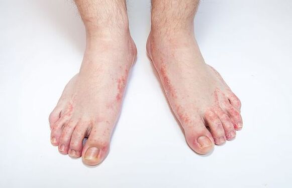
foot fungus(Dermatophytosis,Tinea pedis) is a skin disease of the feet caused by pathogenic or opportunistic fungi.Skin changes on the feet are characterized by peeling, accompanied by itching.When the disease is severe, the skin will be red and swollen, and erosion and deep cracks will appear on the soles of the feet and between the toes, accompanied by pain and difficulty walking.
The code according to the International Classification of Diseases, 10th revision (ICD-10) is B35.3.
The advent of modern antifungal drugs has improved the epidemiological situation, but foot mycosis remains one of the most important problems in dermatology and venereology.The use of certain medications is limited in older adults and people with chronic illnesses.
Prevalence of foot mycosis.According to statistics from the World Health Organization (WHO), about 1/3 of the world's population suffers from fungal diseases, the most common of which is foot mycosis; the incidence rate is increasing year by year.
According to dermatologists, 10-20% of adults suffer from foot fungus; in men, the disease is twice as common as in women and more common in older adults than younger adults.One out of two patients over the age of 70 suffers from foot mycosis, which is associated with an increase in concomitant metabolic and vascular changes (diabetes, varicose veins, etc.).Fungal diseases of the feet are increasingly being detected in children.
Millions of people are currently affected by this disease.Workers in many professions are at risk: miners, athletes and military personnel.
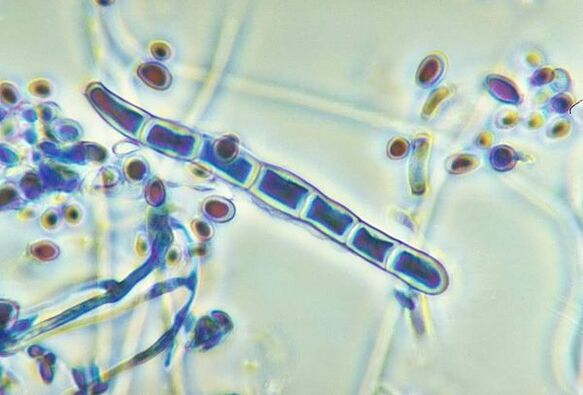
Causes of foot fungus.The most common causes of foot mycosis are dermatophytes: Trichophyton rubrum (90%), Trichophyton mentagrophytes and, less commonly, Epidermophyton.Occasionally, foot mycosis may be caused by fungi of the genus Candida.
Risk factors for foot fungus:
- Exogenous (external): microtrauma to the skin of the feet (calluses, corns), cracks, increased sweating, wearing tight shoes, shoes made of artificial materials, non-observance of personal hygiene rules, irregular foot washing and poor towel drying.
- Endogenous (internal): varicose veins and vegetative vascular dystonia, resulting in insufficient blood supply to the skin of the feet; vitamin deficiencies; taking glucocorticoids, cytostatics, antimicrobials, and estrogen and progesterone drugs, which reduce the body's overall immunity.
Infections with foot fungus can be transmitted directly from a sick person or through contact and household contact (in swimming pools, bathrooms, gyms, through shoes, towels, carpets, etc.).
If you notice similar symptoms, talk to your doctor.Don’t self-medicate – it’s harmful to your health!
Symptoms of foot fungus
The main symptoms of foot fungus:
- itching;
- small cracks;
- erythema;
- peeling;
- bubble;
- Keratosis of the skin;
- Unpleasant and pungent odor;
- Burning, painful sensation.
The first symptoms of foot mycosis are itching and burning in the interdigital folds of the feet, and the skin begins to peel, crack, become red, and show signs of swelling and inflammation.Complications may occur in the form of diaper rash and skin eczema.
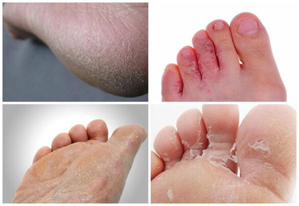
Types of foot fungus:
- Efforts - manifested by moderate itching and congestion (redness) of the skin;
- Acute - skin lesions with severe itching and cracks;
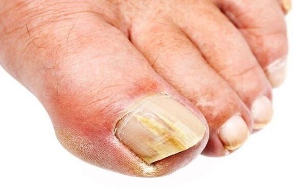
- Onychomycosis (onychomycosis) - manifests as damage to the nail plate, thickening and discoloration of the nail plate;
- Diaper-like - formation of crying area;
- Squamous - appearance of lamellar scales;
- Hyperkeratosis - rash with papules and plaques on the arches of the feet;
- Difficulty sweating - occurs with the development of swelling, oozing areas and blisters.
Pathogenesis of foot mycosis
Skin is the largest organ of the human body, accounting for 15% of body weight.It has several functions, first of all, it protects the body from external factors of physical, chemical and biological nature, prevents water loss and is also involved in thermoregulation.Skin is composed of three layers: epidermis, dermis, and subcutaneous fat.
The epidermis (the outer layer of the skin) is the main barrier for fungi to penetrate the skin.It is a multilayered squamous keratinized epithelium, which is composed of five layers and acts as a barrier.Keratinocytes are the main cells of the epidermis.They contain keratin, which forms the skin's outer layer and gives it elasticity and strength.The keratinocytes of the epidermis are constantly shed.
Dermatophytes produce the enzyme keratinase, which destroys keratin.As a result, the fungus penetrates to the surface of the skin, where it remains.The cell walls of dermatophytes contain mannan, a substance that suppresses local cellular immunity.Due to the effect of mannan, the red fungus T. rubrum can prevent the proliferation of keratinocytes, thereby slowing down the shedding of horny scales on the skin surface and forming a chronic infection process.
Classification and stages of development of foot mycoses
Classification according to pathogens:
- Corneal mycosis (tinea versicolor).
- Dermatophytosis (microsporomycosis, superficial trichophyton, foot mycosis, smooth dermatophytosis, inguinal fold mycosis, onychomycosis).
- Candidiasis (candidiasis of the skin and nails).
- Deep mycoses (blastomycosis, sporotrichosis, chromomycosis).
According to ICD-10 classification:
- B35.1 - Nail fungus.
- B35.2 - Mycoses of the hands.
- B35.3 - Mycoses of the feet.
- B37.2 - Candidiasis of the skin and nails.
Classified by localization:
- Dermatophytosis.
- Pleural mycosis.
- Fungal disease of the hands.
- Podomycoses (scaly, hyperkeratotic, intertriginous, dyshidrotic forms).
- Onychomycosis (distal, superficial, proximal).
According to clinical classification:
- erase formIt manifests as peeling of the III-IV interdigital folds of the feet.Mild peeling may also occur on the soles and sides of the feet.
- interfrictional formIt manifests as congestion in the interdigital folds of the feet, and bubbles may also appear, leading to erosion and cracks.Subjectively, itching and burning sensations were noted.
- Have a form of difficulty sweatingGroups of blisters appear on the skin of the arch and sides of the foot.More often, they appear on healthy skin and then increase in size, coalesce, and form larger, multilocular blisters.When blisters open, erosions form.
- Squamous form of hyperkeratosisIt is characterized by localized or widespread thickening of the stratum corneum on the lateral and plantar surfaces of the foot.The affected areas of skin are covered with small scales that resemble pityriasis.Peeling is especially noticeable in skin folds.Cracks can cause pain when walking.
From a practical perspective, classification by clinic is very convenient for determining further treatment strategies and monitoring patients.
Based on the clinical manifestations of the disease, the cause of the disease can be determined.For example, forms of dyshidrosis often occur with foot mycosis caused by Trichophyton mentagrophytes.The interdigital, squamous hyperkeratosis form is more commonly associated with Trichophyton rubrum, and the chronic and extensive course is characteristic of the opportunistic fungus Candida spp.and Aspergillus spp.
Complications of foot fungus
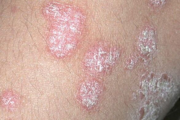
- Allergy to fungi.Under the influence of fungi, polyvalent sensitization is formed, that is, the body becomes more sensitive to the fungal waste products, which are foreign to us and are strong allergens.The body reacts more violently, manifesting itself in various rashes and reactions, allergic chronic conditions, such as skin eczema.The development or exacerbation of conditions such as bronchial asthma, atopic dermatitis, seborrheic dermatitis, and psoriasis is possible.Additionally, a person may be more susceptible to occupational allergy complications and medication intolerances.
- Pyoderma- Pustular dermatoses (cellulitis, lymphangitis, cellulitis, osteomyelitis of the bones of the feet), leading to deep, long-lasting non-healing skin wounds.Pyoderma occurs because bacteria can easily penetrate erosions and cracks in the skin ("gateways to infection").At the same time, the body temperature rises, weakness and discomfort occur, and immediate surgical correction is required.
- Increased viral complicationsThe form of warts develops due to the presence of hyperkeratosis and fissures.The reason is that the protective functions of the skin are violated, making the skin more susceptible to any infection, including viral infections.
- Immunity is generally reducedPatients with medical conditions such as diabetes and varicose veins have impaired microcirculation in the lower extremities.
- The disease spreads to the nails and skin of the hands.When nail fungus occurs, they become deformed and may develop ingrown nails, paronychia (purulent inflammation of the finger tissue), paronychia (inflammation of the periungual folds), and complete loss of the nail plate.
- Quality of life deteriorates.The acute form of foot mycosis can be painful, make it difficult to put on shoes, and, when lymphadenitis is present, can be accompanied by poor general health and fever.
Diagnosis of foot fungus
The diagnosis of foot mycosis is based on the patient's complaint, medical history, clinical findings, and laboratory results.Podiatry is one of the diseases for which laboratory testing is necessary to confirm the clinical diagnosis.
The main methods of diagnosing foot mycosis are microscopy and culture.The material is a skin graft, scraped from a lesion on the skin with a scalpel or glass; less commonly, a tape test is used.
laboratory diagnosisMycoses include microscopic and cultural examination of fungal material.microscopyis a rapid method for diagnosing pathogens and can identify fungal structures within a few hours.Microscopic examination may reveal fungal components in the form of hyphae and spores.The disadvantage of this method is the possibility of obtaining both false positive and false negative results, which depends on many factors: extraction technology, particularities of storage and transportation, etc.
Cultivation methodIt is the most accurate diagnostic method that can identify the fungal type for causative treatment.In preparation for analysis, patients were advised not to use any antifungal agents alone for 1 month.
When administering systemic antifungal therapy, it is recommendedblood biochemistry testLevels of bilirubin, AST, and ALT are determined to meet the needs of monitoring liver and biliary function and to prevent possible complications.
Differential diagnosis of foot mycosis:
- The scaly form is different from psoriasis, eczema, and keratosis.
- Interdigital type is different from impetigo, diaper rash, and candidiasis.
- The dyshidrotic form is distinct from palmoplantar pustulosis.
Treatment of foot fungus
Treatment should be carried out under the supervision of a dermatologist.
The first priority in the fight against foot mycosis is prompt detection, identification and treatment of onychomycosis before it develops, which requires longer and more complex treatment (systemic antifungal therapy).At the same time, it is also important to have effective drugs that match the modern clinical features of foot mycoses.
Before starting to treat a disease, a dermatologist will choose between possible treatment options.In most cases, the medication is applied topically.The basis of treatment is the use of antifungal drugs with different effects.Drugs and medicines that stimulate blood circulation are also used to eliminate the main symptoms:
- Topical antifungal drugs: topical, 1-2 times a day for 4 weeks.
- If the feet are hyperkeratotic, start with an exfoliation treatment: use an azole derivative once a day for 3-4 days, which acts as a keratolytic agent, i.e. removes the rough layer, thus preparing the skin and promoting the penetration of antifungal agents into the dermis.
- If bubbles are present, use Castellani liquid; this solution is applied externally 1-2 times a day for 2-3 days.Then combine the medicines and apply them externally twice a day for 7-10 days.
- For severe itching, antihistamines may be used: Histamine H blockers1-Receptor - ethanolamine derivative 0.001 g, taken orally 2 times a day for 10-15 days.
- Disinfect your shoes once a month until they are completely cured; you can use a spray with the active ingredient undecylenimidyltrimethyl methyl sulfate.
- If the nail plate is affected, oral systemic antifungal treatment is necessary for 3 to 4 months.This therapy requires dermatologist supervision, since self-medication may lead to complications of internal organs (mainly liver, biliary tract, stomach), as well as ineffectiveness of treatment and development of treatment resistance.
It is necessary to treat fungal diseases of the feet, because if the fungus has already settled in the skin, then it will not go anywhere without treatment, which means that the waste products of the fungus will always enter the surrounding tissues and blood, leading to the development of allergies and allergic chronic diseases in the body.
The presence of fungi indicates a decrease in immunity, and skin damaged by fungal diseases is actually unable to perform a protective function.Thus, all conditions are created for an increase in accompanying bacterial infections.
Patients with foot mycosis are active sources of infection for those around them, especially family members, so treatment of this case is an effective means of preventing fungal infection in healthy relatives and surrounding people.
The most favorable environment for foot skin fungal infection is a moist environment, so you need to try to keep the skin on your feet as dry as possible.To do this, you need to wash your feet with soap every night and dry the skin with disposable paper towels, paying special attention to the spaces between the toes.
forecast.prevention
The prognosis of dermatomycoses depends largely on the stage of the disease when treatment is initiated.Therefore, you should not put off seeing your doctor if you notice changes in your skin.Prompt and correct treatment of foot mycosispredictFavorable: Complete recovery from the fungal infection and recovery of the patient.
If left untreated, the fungus can lead to complications that not only deform the nail but also affect the condition of the entire body.
preventionFungal infection:
- Public prevention includes treatment in public places: baths, saunas, swimming pools, showers.Floors, equipment and household items must be disinfected.Personnel and those who frequent public baths, saunas and other places should undergo regular preventive inspections.
Key personal precautions:
- Follow personal hygiene rules when going to public places;
- Avoid damage and continued moisture to the skin and nails of your feet;
- Wear loose, comfortable shoes;
- Avoid contact with infected people.
Secondary personal prevention:
- Maintain foot skin hygiene;
- Disinfect shoes, showers and bathrooms;
- Increase immunity.